Proteins

Proteins, proteomics

Proteins and Proteomics

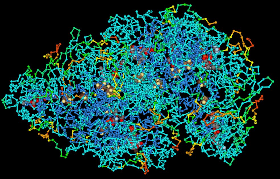
Proteins are complex, macromolecules comprised of amino acids linked by peptide bonds into long chains. The sequence (primary structure) and properties of constituent amino acids generate the 3D conformational structure (tertiary and quaternary structure) that is vital to the biological function of proteins. (click to enlarge image)


Proteins are essential to the structure and biological viability of all living cells and viruses. The cellular proteome is the total cellular protein under a particular set of conditions, while the complete proteome is the sum of all potential proteomes of a cell. Proteomics has become the subject of much research in cell and molecular biology.

Proteins play a number of vital roles as:
a. Enzymes or subunits of enzymes – catalyzing cellular reactions.
b. Structural or mechanical roles – structural components of tissues, components of the cytoskeleton, centrioles, cilia and flagella, microtubules, molecular motors.
c. Intracellular and intercellular signaling functions – ion channels, receptors, membrane pumps.
d. Regulatory proteins or adaptor proteins in genetic transcription, RNA processing, spliceosomes.
e. Products of immune and inflammatory responses that aid in targetting of foreign substances and organisms.
f. Storage and transport of various ligands.
g. The source of essential amino acids.

Almost all natural proteins are encoded by DNA, which is transcribed and processed to yield mRNA, which then serves as a template for translation by ribosomes on the rough endoplasmic reticulum.

Protein roles:
cytoskeletal protein, enzymes, ion channels, latent gene regulatory proteins, receptor proteins, 'signaling' enzymes, signaling proteins, transcription factors
activator,
adaptor protein, amplifier protein, anchoring protein, bifurcation protein, coincidence detector protein, effector protein, messenger protein, modulator protein, relay protein, scaffold protein, transducer protein

Specific proteins/types : § adaptor protein : cAMP receptor binding protein § CARD domains : cofactor § collagen : core histones H2A, H2B, H3, and H4 : CRE-binding protein CREB § C-reactive protein § CRP § domains : elongation factor EF § granulysin : helicases : Helicase II : heterochromatin : histone : HP1 § immunoglobulin isotypes : inducible transcription factors : LexA repressor : mCAT2 receptor : motor proteins § NF-κB : nucleosome : PcG proteins : PCNA § (pentraxins, CRP) § PH family : Polycomb group : proteome : RecA : regulatory proteins : repressor proteins : ribosomes : RPA : serine rich (SR) splicing factors : silencers : Ski7p : small nuclear ribonucleoproteins (snRNPs) : spliceosome : SR (serine rich) splicing factors : trans-acting factors : trithorax group (trxG) : UPF1 UPF2 : upstream transcription factors :

Enzymes ♦ Enzymes : ♦ activation-induced (cytidine) deaminaseadenylate cyclasesAIDAkt : AP endonuclease (Ape1) ♦ ATPasesbondscAMP-dependent protein kinasecyclin-dependent kinases : DNA glycosylase : DNA Ligase I : DNA polymerases : DNA polymerase I : DNA polymerase beta : DNase IVenergetics : exonuclease 1 : exosome : Fen1 : Flap Endonuclease FEN-1Fyn : general transcription factorsGTPases : hOGG1 : hOGG1 oxoG repair : LigIII : MAP kinaseMAPKsMEKMPF : Msh2-Msh3mTOR : MutS, MutL, and MutH : 8-oxoguanine glycosylase : oxoG repair hOGG1 : PCNAPDK1PTENPDK1phosphatasesphospholipasesphosphodiesterasesphosphorylasesPIKKPI3KPKAPKBPKCsprotein kinasesreaction energeticsreceptor tyrosine kinases : RNA polymerase: Replication factor C : reverse transcriptase : ribozymes : RNA polymerase IIserine/threonine kinases : spliceosomal-mediated RNA trans-splicing SMaRT : trans-splicing ribozymesUNG2uracil-DNA glycosylase : UvrD : XRCC1 :

[] Rediscovering Biology - Animations and Images []

Alphabetic links to Glossary items:◦ ABCDEFGHIJKLMNOPQRSTUVWXYZ ◦ o-o animation index o-o diagram index o-o image index o-o micrograph index o-o table index o-o sem/tem index o-o video index .

tags

Labels: , , , , ,

| 0 Guide-Glossary

adaptor protein

An adaptor protein links components in signaling pathways by acting as an accessory to the chief proteins in a signal transduction pathway.

adaptor genes : adaptor proteins : AP-1, AP-2 : Cbl : clathrin : Grb2 : LIME : Nck : RING : SH2, SH3 : thymocyte development : TIR : TLR : Toll-like receptor : ubiquitin-ligases

Adaptor proteins lack intrinsic enzymatic activity, instead they mediate specific protein-protein interactions that drive the formation of protein complexes. The specificity of Toll-like receptor signaling occurs by means of adaptor proteins possessing Toll–interleukin 1 receptor (TIR) domains.

Examples of adaptor proteins include:
Adaptor protein complex 1 (AP-1) and AP-2, which interact with clathrin-coated vesicles located at the trans-Golgi network (TGN) or the plasma membrane. Heterotetramer adaptor protein complexes that function as vesicle coat components and cargo molecule selectors in various membrane trafficking pathways.

Cbl adaptor proteins are RING-type E3 ubiquitin ligases that may be involved in the negative regulation of thymocyte development, targeting its substrate for ubiquitination [ 1 ]. The ubiquitin ligase activity of Cbl, and of its homologue Cbl-b, plays a role in the negative regulation of upstream kinases, such as Lck, Syk and PI3K, in T and B cells

Grb2
IPS-1/MAVS/VISA/Cardif
LIME (Lck-interacting molecule) is a raft-associated adaptor protein that is expressed predominantly in T lymphocytes, and which becomes tyrosine phosphorylated after cross-linking of the CD4 or CD8 coreceptors. Phospho-LIME associates with the Src family kinase Lck and its negative regulator, Csk.

Nck
Src homology = Shc proteins → SH2 recognize phosphotyrosine residues and SH3 domains recognise proline-rich sequences

Genes encoding adaptor proteins include:
GRAP → GRB2-related adaptor protein
GRAP2 → GRB2-related adaptor protein 2
LDLRAP1 → low density lipoprotein receptor adaptor protein 1
NCK1 → NCK adaptor protein 1
NCK2 → NCK adaptor protein 2
NOS1AP → nitric oxide synthase 1 (neuronal) adaptor protein
PIK3AP1 → phosphoinositide-3-kinase adaptor protein 1
SH2B1 → SH2B adaptor protein 1
SH2B2 → SH2B adaptor protein 2
SH2B3 → SH2B adaptor protein 3
SHB → Src homology 2 domain containing adaptor protein B
SLC4A1AP → solute carrier family 4 (anion exchanger), member 1, adaptor protein

tags

Labels: , , , , , , ,

| 0 Guide-Glossary

alpha-1 acid glycoprotein

Alpha-1-acid glycoprotein (AGP) or orosomucoid (ORM) is an acute phase protein synthesized in response to pro-inflammatory cytokines early in the inflammatory response.

AGP gene expression is controlled by a combination of the major regulatory mediators – glucocorticoids and a cytokine network involving IL-1β, TNF-α, IL-6, and IL-6 related cytokines. Human AGP is a 41-43 kDa glycoprotein with single chain of 183 amino acids plus five to six highly sialylated complex-type-N-linked glycans, rendering the molecule heavily glycosylated (45%). Binding affinity and immunomodulatory activities of AGP are mostly dependent on carbohydrate composition.

Alpha-1-acid glycoprotein. Fournier T, Medjoubi-N N, Porquet D. Biochim Biophys Acta. 2000 Oct 18;1482(1-2):157-71.

tags

Labels: , , , ,

| 0 Guide-Glossary

alpha 1-antichymotrypsin

alpha 1-Antichymotrypsin (ACT) is an an acute phase protein synthesized in response to pro-inflammatory cytokines early in the inflammatory response.

ACT is a member of the serine proteinase inhibitor (serpin) family that inhibits neutrophilic proteinases – chymotrypsin, cathepsin G, chymases from mast cells, and elastase – protecting tissue from damage by these proteolytic enzymes. ACT is a glycoprotein found in alpha(1)-globulin region in human serum. Alpha 1-antichymotrypsin contains a reactive centre loop that interacts with cognate proteinases, resulting in loop cleavage and a major conformational change.

As an acute phase protein, ACT is active in the control of immune and inflammatory responses, and acts as a tumor marker. ACT inhibits CTL-mediated lysis (CML).

ACT has been identified as a major constituent of the neurofibrillary plaques associated with Alzheimers disease, where it probably enhances the rate of amyloid-fibril formation. Genetic data also suggests that alpha 1-antichymotrypsin is important in the pathogenesis of Alzheimer's disease.[s]

tags

Labels: , , , , , , , ,

| 0 Guide-Glossary

alpha 1-antitrypsin

Alpha 1-antitrypsin or α1-antitrypsin (A1AT) or alpha-1 proteinase inhibitor (α1-PI) is an an acute phase protein synthesized in response to pro-inflammatory cytokines early in the inflammatory response.

A1AT is a 52 kDa prototypical serine protease inhibitor (serpin) that protects against enzymes released by inflammatory cells, particularly elastase, which is released from neutrophilic granules. A1AT forms covalent bonds with both elastase and trypsin, irreversibly inactivating these proteolytic enzymes.

Alpha 1-antitrypsin deficiency is a hereditary disorder in which inability to inactivate elastase and trypsin allows inflammatory tissue breakdown, causing pulmonary emphysema and hepatic cirrhosis in severe cases.

tags

Labels: , , , , ,

| 0 Guide-Glossary

alpha 2-macroglobulin

Alpha-2 macroglobulin (A2M), α-2 macroglobulin is an an acute phase protein synthesized in response to pro-inflammatory cytokines early in the inflammatory response.

A2M is a large plasma protein produced by the liver, and is a major component of the alpha-2 band in protein electrophoresis. A2M interacts and captures virtually any proteinase whether self or foreign, suggesting a function as a unique "panproteinase inhibitor." A2M also removes the active forms of the gelatinase (MMP-2 and MMP-9) from the circulation, binding to scavenger receptors on the phagocytes. In the mechanism termed "clearance of activated alpha 2-macroglobulin", ACT undergoes Ca(2+)-dependent binding to a member of the low-density lipoprotein receptor supergene family that mediates cellular uptake by endocytosis and delivery to endosomes and lysosomes. Thus, the peptide binding function of A2M allows targeting of biologically active peptides to different cell types expressing the A2M receptor. Complexes internalized through this binding may be dispatched into different pathways of endocytic/lysosomal pathways in a cell type-specific manner.[r]

A2M ratios are increased in nephrotic syndrome when the kidneys lose smaller plasma proteins (proteinuria). A common variant of α2-macroglobulin is associated with increased risk of Alzheimers disease.

 Scavenger Receptors

tags

Labels: , , , , , , , , ,

| 0 Guide-Glossary

CARD domains

Caspase recruitment domains, or CARD domains are found in many proteins, particularly those that evoke inflammatory responses or apoptosis. Proteins with CARD domains include caspases, helicases, kinases, mitochondrial proteins, and other cytoplasmic factors.

CARD domains mediate the formation of large protein complexes through direct interactions between individual caspase recruitment domains. The domains belong to a subclass of protein motif termed the 'death fold', which comprises six to seven antiparallel alpha helices with a hydrophobic core and an outer face composed of charged residues, and which is conserved at least as far as the ced-3 and ced-4 genes in C. elegans. Other 'death fold' motifs in this class are found in the pyrin domain (PYD), death domain (DD), and death effector domain (DED). All function primarily in regulation of apoptosis and inflammatory responses.

The NACHT–leucine-rich repeat (NLR) protein, Ipaf-1 features an N-terminal CARD domain, a nucleotide-binding domain, and C-terminal leucine-rich repeats (LRRs), homologous to those found in Toll-like receptors. Ipaf-1 has a primary role in regulation of proteolytic processing of pro-IL-1β and pro-IL-18 into their mature forms by way of association in a large complex known as the inflammasome.

Ipaf-1 activation by the intracellular bacterium S. typhimurium or other stress signals, causes Ipaf-1 to recruit a CARD-containing adapter termed ASC and caspase-1. This ASC•caspase-1 complex activates caspase-1 and leads to IL-1β and IL-18 maturation.

CARD proteins also participate in recognition of intracellular dsRNA found in a number of viral genomes, including the para-, orthomyxoviridae, and rhabdoviridae. Unlike NLRs, these RIG-I and MDA5 proteins contain twin N-terminal CARD domains plus C-terminal RNA helicase domains that directly interact with and process the double-stranded viral RNA. This processing renders the CARD domains available for interaction with the CARD motif of IPS-1/MAVS/VISA/Cardif, which is a mitochondrion-anchored downstream adaptor.

Because CARD proteins function as regulators of inflammation, the constitutive activation of certain CARD proteins might play a causative role in some inflammatory syndromes.

tags

Labels: , , , ,

| 0 Guide-Glossary

coagulation factors

Coagulation factors participate in the coagulation cascade, which is initiated within 20 seconds of an injury to the endothelial cells that line blood vessels (primary hemostasis, acute phase reaction).

Coagulation factors are synthesized by the liver and their production increases in response to pro-inflammatory cytokines released in the acute phase early in the inflammatory response, in particular:
● factor VIII
● fibrinogen
● plasminogen
● prothrombin
● von Willebrand factor

tags

Labels: , , ,

| 0 Guide-Glossary

collagen

Collagen is the chief structural protein of connective tissue in mammals, located mostly in the extracellular matrix. Collagen comprises long chains containing unusually large amounts of proline, together with glycine residues at approximately every third position. Collagen also contains two uncommon derivative amino acids, hydroxylysine and hydroxyproline (Hyp), where hydroxylysines in some collagens have attached disaccharides.

The tropocollagen subunit is a rod (300 nm x 1.5 nm) comprising three polypeptide strands, each of which is a left-handed helix (not an α helix). Collagen self-assembles, and the precise alignment of individual collagen molecules produces a characteristic pattern of light and dark bands across the fibril on electron-microscopy [] ce-tem collagen fibril [].

Alphabetic links to Glossary items:◦ ABCDEFGHIJKLMNOPQRSTUVWXYZ ◦ o-o animation index o-o diagram index o-o image index o-o micrograph index o-o table index o-o sem/tem index o-o video index .

Companion Sites : Gray Sites α Abiogenesis & EvolutionAlgorithms of Evolution ¤ CancerCell Biology ~ Chemistry of Life Ω Complex Systems ººº CyanobacteriaEnzymesEvo Devo Ơ Fat ф Immunology »» Mechanisms of EvolutionMolecular Biology ** Origin of LifePaleogeology § Protein ¿ Refuting ID ǒ Serial Endosymbiosis ζζ SleepStromatolitesTaxonomy PhylogenyTissue ۰۰ Virus • White Sites  Diagrams & Tables æ Eubacteria & Archaea φ MoleculeMolecular Paths ››› PathwaysPhotosynthesis • Black Sites Ǒ Endosymbiosis ō Organics

tags

Labels: , , ,

| 0 Guide-Glossary

C-reactive protein

C-reactive protein is an acute phase protein produced by the liver within 6 hours of an acute inflammatory stimulus. Levels peak after about 50 hours.

CRP is a pentraxin protein that exhibits calcium dependant ligand binding and a distinctive flattened β-jellyroll structure. (left - image - click to enlarge) CRP is so-named because it reacts with the somatic C polysaccharide of the bacterium Streptococcus pneumoniae. Serum amyloid P component is another pentraxin, acute phase protein.

CRP binds specifically to phosphocholine moieties, conferring a host-defensive role on CRP since phosphocholine is a component of microbial polysaccharides. CRP also binds to ligands exposed on damaged cells.

CRP-phosphocholine-binding:
● activates the classical complement pathway
● acts as an opsonin ligand for phagocytosis
● neutralizes the pro-inflammatory platelet-activating factor (PAF)
● down-regulates polymorphs
CRP
● increases production of tissue-factors by monocytes
● activates smooth muscle K+ ion channels (vasodilator)
● delays apoptosis of neutrophils when the pentameric structure is lost and the molecule exists as a monomer (mCRP)

CRP is mildly elevated in: systemic lupus erythematosus, systemic sclerosis, sermatomyositis, ulcerative colitis, leukaemia, and graft-versus-host disease (GVHD).

The C-reactive protein medical test measures levels of CRP to assess acute inflammation. An association has been demonstrated between sudden cardiac death, peripheral arterial disease and hs-CRP, so serum CRP levels may correlate with cardiovascular disease risk.

tags

Labels: , , , , , , , , ,

| 0 Guide-Glossary

domains

A binding domain is that sequence of amino acids in a protein/protein family to which a specific ligand binds. As such, domains are vital to a protein's or enzyme's function. A structural domain is a self-stabilizing structural element that may fold independently of the rest of the protein chain.

Specific examples of domains:
cadherin repeats
carbohydrate-recognition domain (CRD)
caspase recruiting domains, CARD domains
C-lectin domain (CRD)
C-type-lectin-like domain (CTLD)
death domain (DD), death effector domain (DED) binds adaptor protein FADD (Fas-Associated Death Domain)
EF-hand domains
kringle domains
pleckstrin homology (PH) domain family
SH2 domain - Src homology 2 domain = p-Tyr recognition domains
zinc finger DNA binding domains

Others (on Wiki) Arginine Finger, Armadillo repeats, Basic Leucine zipper domain (bZIP domain), Phosphotyrosine-binding domain (PTB)

Labels: , , , , , , , , , , ,

| 0 Guide-Glossary

EF hand domains

EF-hand domains comprise two helices that are arranged roughly perpendicular to each other and connected by a loop. Most EF-hand units are paired to form a compact lobe, and often bind calcium ions with ligands located in the loop that connects the helices.

EF-hand Ca(2+)-binding proteins participate both in modulation of calcium signals and in direct transduction of an ionic signal into downstream biochemical events. The biochemical repetoire of EF-hand proteins is correlated with differential response to conformational changes caused by Ca(2+) binding. For example, calmodulin and calbindin have homologous EF-hand domains, yet they respond to calcium binding differently.[pm]

Labels:

| 0 Guide-Glossary

ferritin

Ferritin is an an acute phase protein synthesized in response to pro-inflammatory cytokines early in the inflammatory response.

Ferritin is the chief iron storage protein for both eukaryotes and prokaryotes, and is termed apoferritin when not combined with Fe(3+) ions. The globular ferritin protein complex comprises 24 subunits – hetero-oligomers of light (L) and heavy (H) chains encoded by homologous genes (vertebrates), H chains only in bacteria and plants. Free Fe is toxic, so cells employ ferritin molecules or aggregates (hemosiderin) to form protective Fe-protein complexes. Each ferritin molecule complexes around 4500 (Fe3+)ions.

Ferritin is found in most tissues, particularly in the bone marrow and reticuloendothelial system (or mononuclear phagocytic system). Serum ferritin levels are proportional to iron storage levels in humans and are employed as a part of the work-up for anemia, particurlarly iron-deficiency anemia. However, ferritin levels are increased by acute inflammation, hemochromatosis, malignancy, hyperthyroidism, Still's Disease, or hepatic disease (necrotic hepatocytes), so ferritin levels may be misleading when anemia is concomitant with these conditions.

tags

Labels: , , , , ,

| 0 Guide-Glossary

fibronectin

Fibronectin (FN) is a high molecular weight, multidomain glycoprotein, comprising about 5% by weight of carbohydrate.

Fibronectin exhibits diverse recognition functions located on distinct fragments or domains, so FN can interact with a variety of macromolecules including/on :
cytoskeleton
extracellular matrixcollagen, glycosaminoglycans, proteoglycans, tenascin, fibulin and thrombospondin
● circulating coagulation factors – Fn is covalently incorporated into fibrin clots through the transglutaminase action of coagulation factor XIII, improving fibroblast adhesion
fibrinolytic system
acute phase proteins
complement system
cell-surface receptors on a variety of cells including fibroblasts, neurons, phagocytes and bacteria – integrins (through RGD tripeptide)
● itself, forming fibrillar entities
● small molecules such as gangliosides, sugars, and Ca ions.

Fibronectin (FN) participates in tissue repair, embryogenesis, blood clotting, and cell migration/adhesion. Cells of most tissues synthesize fibronectin. Soluble fibronectin is produced by hepatocytes and circulates, in its disulfide-bonded dimeric form, in the plasma. The soluble protomer is a compact, flexible dimer that can be converted into an insoluble, fibrillar network. The soluble-to-fibrillar conversion is a highly regulated process involving integrins and possibly other cell-surface receptors [ref] including uPAR (urokinase-type plasminogen activator receptor) [ref] and a cell-surface proteoglycan [ref].[s]

The insoluble fibronectin dimer is synthesized by fibroblasts, chondrocytes, endothelial cells, macrophages, as well as certain epithelial cells. Electron microscopic analyses of natural thin fibrils (5-18nm diameter), made by fibroblasts in culture, clearly indicate an ordered arrangement and suggest a model in which extended protomers (130nm long) are arranged end-to-end with an overlap of about 14 nm [ref]. As an extracellular adhesion molecule, FN binds to integrins and participates in wound healing.

Cell-surface receptors or fibrinogen, collagen and fibrin (as extracellular matrix proteins) facilitate the adherence of microorganisms to host tissues [ref]. The Hep-2 domain of fibronectin interacts with envelope glycoproteins on some retroviruses. Fibronectin is able to bind both the virus and cell-surface receptors, concentrating viruses on the surface of the cells, enhancing viral uptake by cells.

The structural isoforms of fibronectin arise from alternative splicing of a single gene, and possess a variable region plus three types of repeated internal regions (homologous, repeating modules I, II and III) +/- disulfide bonds.

[more] [] The Type I module of fibronectin [] The Type II module (F2) [] segment of fibronectin , four Type III modules []

tags

Labels: , , , , , , , , ,

| 0 Guide-Glossary

granulysin

Granulysin is a 74-aa basic protein released from human cytolytic T lymphocytes (CTL, Tc) and natural killer cells (NK).

'The five-helical bundle of granulysin resembles other "saposin folds" (such as NK-lysin). Positive charges distribute in a ring around the granulysin molecule, and one face has net positive charge. Sulfate ions bind near the segment of the molecule identified as most membrane-lytic and of highest hydrophobic moment. The ion locations may indicate granulysin's orientation of initial approach towards the membrane. The crystal packing reveals one way to pack a sheet of granulysin molecules at the cell surface for a concerted lysis effort. The energy of binding granulysin charges to the bacterial membrane could drive the subsequent lytic processes. The loosely packed core facilitates a hinge or scissors motion towards exposure of hydrophobic surface that we propose tunnels the granulysin into the fracturing target membrane.'[pm] Granulysin crystal structure and a structure-derived lytic mechanism. Anderson DH, Sawaya MR, Cascio D, Ernst W, Modlin R, Krensky A, Eisenberg D. J Mol Biol. 2003 Jan 10;325(2):355-65.

tags

Labels: , , ,

| 0 Guide-Glossary

haptoglobins

Haptoglobins (Hp) are acute phase proteins synthesized in response to pro-inflammatory cytokines early in the inflammatory response.

Haptoglobins are alpha-2-globulins that remove free hemoglobin from plasma by forming a stable complex that aids in recycling of heme iron.

Haptoglobins are elevated in inflammation, malignancies (particularly with metastases to bone), trauma, surgery, steroid or androgen therapy, and diabetes.

tags

Labels: , , , ,

| 0 Guide-Glossary

immunoglobulin isotypes

The immunoglobulin superfamily comprises an evolutionarily ancient, widely expressed, constitutive or long-term up-regulated family of glycoproteins involved in cellular adhesion, signaling, and the adaptive immune response.

Immunoglobulins (left - click to enlarge) comprise two heavy (H) and two light-chain (L) protein subunits, each of which folds into domains (4 on heavy, 2 on light). These adhesion sites or domains contain one or more folds of 60 to 100 amino acids.

Depending upon the character of the heavy chain, immunoglobulins are divided into five classes – IgG, IgD, IgE, IgA, IgM – that are expressed in different tissues (detail). The classes are further subdivided into isotypes, which have different properties in terms of complement fixation and binding affinity to immunoglobulin (Ig) receptors.

Immunoglobulins can be divided into five different classes, based on differences in the amino acid sequences in the constant region of the heavy chains. All immunoglobulins within a given class will have very similar heavy chain constant regions. These differences can be detected by sequence studies or more commonly by serological means (i.e. by the use of antibodies directed to these differences).
1. IgG - γ - Gamma heavy chains
2. IgM - μ - Mu heavy chains
3. IgA - α - Alpha heavy chains
4. IgD - δ - Delta heavy chains
5. IgE - ε - Epsilon heavy chains

As adhesion-signaling molecules, immunoglobulins act as B-cell receptors (BCR) that recognize (ligate) specific antigens. The B cell receptor (BCR) is an integral membrane protein complex that is composed of two Ig heavy chains, two Ig light chains and two heterodimers of Ig-α and Ig-β. Antigens bind with greatest affinity to their cognate Ig-antibody, and hence to activated the BCR of greatest affinity.

Antigen-activated B cells proliferate and differentiate into memory B cells or plasma cells. B cell development is a tightly controlled process in which over 75% of the developing cells become apoptotic because of inappropriate immunoglobulin gene rearrangements or recognition of self antigens by Igs. Copious amounts of monoclonal antibodies are synthesized by antigen-stimulated B cell-derived plasma cells, while memory B cells remain primed for rapid activation by a repeat encounter with the initial, diffentiation activating antigen.

The enormous diversity of immunoglobulin molecules is attributable to VDJ recombination, while secondary antibody diversification is engineered by AID-induced/UNG2 assisted somatic hypermutation (SHM) or class-switch recombination (CSR), and AID-induced gene-conversion (GC). Affinity maturation is achieved are somatic hypermutation and clonal selection, which together ensure that repeated exposures to the same antigen will provoke greater antibody ligating affinity in the antibody secreted by successive generations of plasma cells.

tags

Labels: , , , , , , ,

| 0 Guide-Glossary

kringle domains

Kringle domains, named for Scandinavian pastries, are conserved sequences that fold into large loops (stabilized by 3 disulfide linkages) the conformation of which is defined by hydrogen bonds and small pieces of anti-parallel β-sheet. Plasminogen-like kringles display affinity for free lysine and for lysine-containing peptides.

Kringle domains are found in several serine proteases, including prothrombin (with two kringle domains) and urokinase-type plasminogen activator, in ROR-like receptors, and they participate in protein-protein interactions with blood coagulation factors.

Urokinase-type plasminogen activator is a strong plasminogen activator which specifically cleaves the proenzyme/zymogen plasminogen to form the active enzyme plasmin.[s] Ror-family RTKs are characterized by the intracellular tyrosine kinase domains, highly related to those of the Trk-family RTKs, and by the extracellular Frizzled-like cysteine-rich domains (CRDs) and Kringle domains.[pm]

[] plasminogen, rotate human plasminogen, primary structure []

tags

Labels: , , ,

| 0 Guide-Glossary

mannose-binding protein

Mannose binding protein (MBP) or mannan-binding lectin (MBL) is a member of the collectin family, and is a C−type serum lectin that participates in the innate inflammatory response in defense against a variety of bacterial, fungal and viral pathogens.

acute phase : CD45 : collectins : domains CRD vs CTLD vs CARD : evolution : ficolins : inflammatory response : lectin pathway of complement activation : MBL-MASP : mutations : receptor : T-cells

MBP is an acute phase protein synthesized in response to pro-inflammatory cytokines early in the inflammatory response. The receptor that recognizes CRD-carrying MBP, and structurally-related complement or lectin opsonins (collectins), is widely distributed on leukocytes, platelets, and endothelial cells.

MBP initiates the lectin branch (MBL-MASP) of the complement cascade by binding to sugars on the surfaces of microorganisms, activating two MBP-associated serine proteases (MASP-1 and MASP-2). MBP is able to specifically recognize cell surface CD45 on immature, but not mature T cells.

The lectin pathway (MBL - MASP) is homologous to the classical pathway, but utilizes opsonin, mannan-binding lectin (MBL, MBP) and ficolins rather than C1q. Binding of mannan-binding lectin to mannose residues on the pathogen's surface activates the MBL-associated serine proteases, MASP-1, MASP-2, MASP-3, which cleave C4 into C4b and C2 into C2b. As in the classical pathway, C4b and C2b bind to form the C4b•C2b C3 convertase. Ficolins are homologous to MBL and function through MASPs. Diversified ficolins are of particular importance in invertebrates, which lack the adaptive immune response that evolved some 500 million years ago in jawed vertebrates. [] C-lectin phylogenetic tree, Immune branch, Snake toxins branch, Calcium-carbohydrate binding branch []

Mutations in the gene encoding mannose binding protein may result in defective opsonization. MBP is a 53 kDa homomultimeric trimer [] ribbon structure MBP [] Human mannose binding protein CRD [] space-fill structure hMBP [] superimposition of CRD/CTLD domains of mannose binding protein C (orange), IX/X binding protein (red), lithostathine (green), human CD94 (purple) []

MBP carries the CRD domain, whose major function is calcium-dependent recognition of oligosaccharides at cell surfaces (carbohydrate-recognition domain (CRD not CARD, which is caspase recruitment domain). The C-type-lectin-like domain (CTLD) is located in a large variety of proteins and is similar to the CRD domain on C- lectins (C-lectin domain). Although CTLDs are similar to CRDs of C-lectins in sequence and structure, they serve different functions.

▲ф ф activation ф activation complement cascade : acute phase ф acute phase : CD45 : collectins ф complement system : domains CRD vs CTLD vs CARD : evolution »» Evolution : ficolins ф humoral immunity ф immune cytokines ф immune response ф inflammatory response : inflammatory response : lectin pathway of complement activation : MBL-MASP : mutations »» Mutation ф pathogens : receptor ф receptors : T-cells ф T cells ▲ф

Top

tags

Labels: , , , , , , , , ,

| 0 Guide-Glossary

modulator proteins

Modulator proteins regulate the activity of other proteins.

Calmodulin (Ca2+-regulated modulator protein, CaM) is a ubiquitous eukaryotic intracellular calcium receptor that regulates the biological activities of many cellular proteins and transmembrane ion transporters.

Calmodulin possess four EF-hand domains that alter conformation upon binding calcium ions. Calcium ions bind to the EF-loop region, shifting the relative positions of the EF-alpha helices. (The other group of proteins that depend on calcium ions for their function belong to the annexin family, which bind calcium and phospholipids). In the absence of calcium, the α-helices in the EF-hand motif of calmodulin are in the closed conformation, almost parallel to each other. When the intracellular calcium level rises, Ca2+ ions bind to calmodulin, causing calmodulin to open into a dumbell shape and increasing CaM's binding affinity. (Nine of CaM's 148 aa residues are methionines, and 8 of these 9 Met are directly involved in binding to all target peptides.) The open conformation Ca2+-calmodulin complex binds target proteins, initiating various signaling cascades. Over a hundred proteins are known to bind calmodulin (above left are three). Calmodulin is essential for cell-cycle progression through mitosis. []molecule of month[]

Labels: , , , , , , , ,

| 0 Guide-Glossary

molecular motors

Molecular motors are proteins or protein complexes that transform chemical energy – derived from ATP hydrolysis – into mechanical work. Molecular motor proteins are of interest both in molecular biology and nanotechnology.

Motors are classified as rotatory or translatory. Eukaryotic translatory proteins translocate materials along filaments (actin filaments, microtubules) and play a role in muscular contraction, cell division, and cellular material transport.

Rotatory:
ATP synthase is the final enzyme of oxidative phosporylation, and the F1Fo ATP synthase is the commonest chemi-mechanical motor found in nature.
topoisomerase operates to unwind DNA molecules – it severs one strand of the double helix, allowing it to relax, and then repairs the cleaved location.

Translatory:
● myosin, which moves along actin microfilaments,
kinesin and dynein, which move along microtubules (tubulin filaments).

Џ Video explaining operation of the F1-F0 ATPase (Harvard U) Џ

[] ATP synthase [] kinesin []

. DNA packing in bacteriophage ø29 (phi-29) . Dynein . Kinesin . Myosin . molecular motors .

Labels: , , , , , ,

| 0 Guide-Glossary

NF-κB

NF-κB, Nuclear Factor kappa Bs, are ubiquitous transcription factors involved in responses to cellular stressors such as cytokines, free radicals, UV irradiation, bacterial antigens, and viral antigens.

action of IκB : actions of NF-κB : family : free NF-κB : IκB : IkappaBalpha : IκB kinase : IKK : NF-κB family : NF-κB/rel : tumors and NF-κB : v-Rel

Members of the NF-κB family are classified as NF-κB/Rel proteins because they display structural homology to the retroviral oncoprotein v-Rel, and include mammalian members:
● N-terminal homology with v-Rel
___ ● NF-κB1 (p50)
___ ● NF-κB2 (p52)
_plus trans-activation domain in C-termini
____ ● RelA (p65)
____ ● RelB
____ ● c-Rel

[] 3D transcription factor NFkappaB -helix-loop-helix []

NF-κB1 and NF-κB2 are synthesized as large precursors (p105, and p100) that undergo selective degradation of their C-terminal region by the ubiquitin/proteasome pathway to generate the mature NF-κB subunits ( p50, and p52 respectively). Degradation of p100 to p52 is tightly regulated process, whereas p50 is generated by constitutive processing of p105. RelA, RelB, and c-Rel contain trans-activation domains at their C-termini. (p52 is not to be confused with the tumor suppressor gene p53.)

Inhibitor of kappa B (IκB, IkappaBalpha) inactivates NF-κB by sequestering NF-κB dimers within the cytoplasm. Activation of IκB kinase (IKK) by stress signals stimulates phosphorylation of two serine residues in IκB's regulatory domain, targetting the IκB molecules for ubiquitin/proteasome degradation, and releasing NF-κB from inhibition.

Free NF-κB translocates to the nucleus where it binds to specific κB sequences in DNA, initiating transcription of related genes, including those for immunoreceptors, cytokines, and its own inhibitor, IκB. Physiological activities mediated by NF-κB include cellular proliferation, and inflammatory, immune, and cellular survival responses. [] signaling pathways []

Many tumor types have chronically active, dysregulated NF-κB, resulting from:
mutations in genes encoding the NF-κB transcription factors themselves, or
mutations in genes that control NF-κB activity

tags

Labels: , , , , , , , , , ,

| 0 Guide-Glossary

PDZ domains

PDZ domains are protein interaction domains found in diverse signaling proteins in bacteria, yeasts, plants, insects and vertebrates.

They comprise ~80-90 residues folded into 6 β-strands and 2 α-helices (image), can occur in one or multiple copies, and are nearly always found in cytoplasmic proteins. The interaction between a PDZ domain and its target is usually constitutive, and the domains bind either the carboxyl-terminal sequences of proteins or internal peptide sequences. PDZ domains bind to the C-terminal 4-5 residues of their target proteins, which are frequently transmembrane receptors such as GPCRs, or ion channels. (image, image, in synapse, diagram)

The consensus binding sequence contains a hydrophobic residue, which is commonly valine or isoleucine, at the very C-terminus. Residues at the -2 and -3 positions determine specificity. PDZ domains can also heterodimerize with the PDZ domains of different proteins, potentially regulating intracellular signaling. Several PDZ domains including those of syntenin, CASK, FAP, and Tiam1 can bind to the phosphoinositide PIP2. Such PIP2-PDZ domain binding is believed to control the association of PDZ domain-containing proteins with the plasma membrane.

Labels: , , , , , , , ,

| 0 Guide-Glossary

PH family

The pleckstrin homology (PH) domain family includes proteins with the PH domain, which is a 100-120 aa module found in a multitude of intracellullar proteins with widepread functions. Many proteins containing PH domains participate in signaling cascades. Other classes of proteins carry a PH domain, including cytoskeletal proteins such as spectrin. [w]

The inositol phospolipids are particularly effective in specific binding to PH domains of cellular proteins. Phosphatidylinositol is an important lipid, both as a participant in essential metabolic processes and as a key membrane constituent. In addition to their role as negatively charged building blocks of membranes, the inositol phospolipids (including the phosphatidylinositol phosphates, or 'polyphosphoinositides') appear to have crucial roles in interfacial binding of proteins and in the regulation of proteins at the cell interface.

Alphabetic links to Glossary items:◦ ABCDEFGHIJKLMNOPQRSTUVWXYZ ◦ o-o animation index o-o diagram index o-o image index o-o micrograph index o-o table index o-o sem/tem index o-o video index .

Companion Sites : Gray Sites α Abiogenesis & EvolutionAlgorithms of Evolution ¤ CancerCell Biology ~ Chemistry of Life Ω Complex Systems ººº CyanobacteriaEnzymesEvo Devo Ơ Fat ф Immunology »» Mechanisms of EvolutionMolecular Biology ** Origin of LifePaleogeology § Protein ¿ Refuting ID ǒ Serial Endosymbiosis ζζ SleepStromatolitesTaxonomy PhylogenyTissue ۰۰ Virus • White Sites  Diagrams & Tables æ Eubacteria & Archaea φ MoleculeMolecular Paths ››› PathwaysPhotosynthesis • Black Sites Ǒ Endosymbiosis ō Organics

tags

Labels: , , , , , , ,

| 0 Guide-Glossary

scaffold proteins

Scaffold proteins either hold protein kinases in a latent state close to their activating cell-surface receptors, or facilitate flow of activation from one kinase to the next kinase in a signaling cascade.

The A kinase anchoring proteins (AKAPs) are prototypical scaffold proteins that organize the protein kinases and phosphatases that regulate serine/threonine phosphorylation: 'Multiprotein signaling networks create focal points of enzyme activity that disseminate the intracellular action of many hormones and neurotransmitters. Accordingly, the spatio-temporal activation of protein kinases and phosphatases is an important factor in controlling where and when phosphorylation events occur. Anchoring proteins provide a molecular framework that orients these enzymes towards selected substrates. A-kinase anchoring proteins (AKAPs) are signal-organizing molecules that compartmentalize the cAMP dependent protein kinase, phosphodiesterases, and a variety of enzymes that are regulated by second-messengers.'[s].

In an example of scaffold mediated assembly of signaling pathways, Jip1 has separate binding sites for Jnk, and for the upstream kinases MKK7 (MAPKK), MLK3 (MAPK), and HPK1, enabling Jip1 to act as a scaffold for the mammalian Jnk MAPK cascade. Jip1 also possesses SH3 and PTB domains that can tether the complex to additional proteins participating in upstream activation or localization. B

ERK MAPK docks to target proteins and phosphorylates the substrate, Rsk1.

The regulatory cyclin A subunit of CDK2 binds substrates with a conserved RXL motif, such as p107.

The AKAP protein Yotiao binds the NMDA receptor, inactive PKA, and active PP1. Thus, Yotiao brings together components that repress resting NMDA receptor and enhance channel activation.[s]

Labels: , , , , , , , , ,

| 0 Guide-Glossary

signaling proteins

Proteins and other molecules serve one or more of several types of function in signaling networks:
cell surface
_ion channels
_receptor proteins
__latent gene regulatory proteins
intracellular
_'signaling' enzymes
_adaptor proteins
_amplifier proteins
_anchoring proteins
_bifurcation proteins
_coincidence detectors
_effector proteins
_mediator molecules
_messenger proteins
_modulator proteins
_relay proteins
_scaffold proteins
_second messengers
_transducer proteins

Adaptor proteins link the components of signaling pathways by acting as accessories to the chief proteins in a signal transduction pathway.

Amplifier proteins are usually either enzymes or ion channels. Amplifiers increase the received signal by producing large amounts of small intracellular second messengers or by activating large numbers of downstream intracellular signaling proteins. Multiple amplification steps in a relay chain are often referred to as a signaling cascade.

Anchor proteins provide a molecular framework that orients these enzymes towards selected substrates. A-kinase anchoring proteins (AKAPs) are signal-organizing, scaffold proteins that compartmentalize the cAMP dependent protein kinase, phosphodiesterases, and a variety of enzymes that are regulated by second-messengers.

Bifurcation proteins spread the signal from one signaling pathway to others.

Coincidence detectors are signaling enzymes that are only activated by several different signals occurring together. For example, cAMP-generating adenylyl (adenylate) cyclases are modulated by G-proteins, forskolin, Ca2+/calmodulin, and other class-specific substrates.

Non-genetic classes of regulatory proteins include those target, effector proteins that are involved in special cellular functions such as signaling as receptor proteins and pumps, adhesion, chemotaxis, cellular transport and active transport, and metabolic regulation, including enzymatic action and protein degradation. Effector molecules bring about regulation by binding other molecules, and genetic effector molecules can participate in the regulation of gene expression. Modulator molecules bind to a regulatory sites during allosteric modulation, where effectors act as activators or inhibitors. (Allosteric proteins have an active (catalytic) site and an allosteric (effector) site.) Effector proteins regulate the activity of other proteins.

Integrator proteins integrate signals from two or more signaling pathways and relay the signal onward along a single pathway.

Latent gene regulatory proteins are activated by receptors at the cell surface and migrate to the nucleus, where they stimulate gene transcription.

Mediators act as molecular go-betweens in signaling cascades. Second messengers are small, readily diffusible molecules that operate in signal transduction (calcium ions, cAMP, cGMP, ceramide, diacylglycerol, inositol-1,4,5-trisphosphate).

Messenger proteins carry the signal from one part of the cell to another. For example, messengers often carry signals from the cytosol to the nucleus.

Modulator proteins regulate the activity of other proteins. Calmodulin (Ca2+-regulated modulator protein, CaM) is a ubiquitous eukaryotic intracellular calcium receptor that regulates the biological activities of many cellular proteins and transmembrane ion transporters. The Ca2+•calmodulin complex binds target proteins, initiating various signaling cascades.

Relay proteins pass the message to the next signaling component in the chain without otherwise participating.

Scaffold proteins either hold protein kinases in a latent state close to their activating cell-surface receptors, or facilitate flow of activation from one kinase to the next kinase in a signaling cascade. The A kinase anchoring proteins (AKAPs) are prototypical anchor/scaffold proteins that organize the protein kinases and phosphatases that regulate serine/threonine phosphorylation.

Transducer proteins convert the signal from one form to another. Signaling enzymes that produce cyclic AMP, for example, both convert the received signal and amplify it (transducer and amplifier).

'Signaling' domains are found in intracellular proteins that are involved in signaling cascades:
EF hand domains
PDZ domains
pleckstrin homology (PH) domain family
SH2 domains

Cell-surface adhesion interactions and inflammatory responses employ specialized domains:
cadherin repeats
carbohydrate-recognition domain (CRD)
caspase recruiting domains, CARD domains
C-lectin domain (CRD)
C-type-lectin-like domain (CTLD)
death domain (DD), death effector domain (DED) binds adaptor protein FADD (Fas-Associated Death Domain)
zinc finger DNA binding domains

Labels: , , , , , , , , , , , , , ,

| 0 Guide-Glossary

Science leads to Atheism

Purists say that science does not lead to atheism, and, in a strict sense, this may be the case. However, most scientists, particularly biological scientists, are agnostic or atheistic. Why? The findings of science provide better explanations than simplistic goddidit claims for supernatural metaphysics and miracles. Even though scientists did not set out to debunk the supernatural, scientific findings have pushed supernatural explanations further and further out of explanatory contention.
Join the best atheist themed blogroll!A-Deistic
Adeistic
Black Out
cosmic
Eine Kleine Nattermusing
eMusings
eVolition
Galaria
Godborygmi
Godspell Follies
Gray Matters
MetaThoughts
Mimble Wimble
Naturalism
BLogodaedaly
palimpsest
Salient
Science of Evolution
Sechuam
Sintheist
Tabula Flexuosa
The Scarlet A
Weltschauung

| 0 Guide-Glossary

affinities

Associated science sites • Abiogenesis and EvolutionAlgorithms of EvolutionArchea EubacteriaCancerCell BiologyComplex SystemsCyanobacteriaDiagrams TablesEndosymbiosisEnzymesEvo DevoEvolution in ActionFatGeologyGlossaryImmunologyLife ChemistryMedical ScienceMechanisms of EvolutionMoleculeMolecular BiologyMolecular PathsNeurosciencesOrganicsOrigin of LifePaleogeologyPathwaysPhotosynthesisProteinReceptorRocks & MineralsSETSignalingSleepStem & Progenitor CellsStromatolitesTaxonomy PhylogenyTissueVirus • And some philosophy/general interest sites • A-DeisticAdeisticBlack OutcosmicEine Kleine NattermusingeMusingseVolitionGalariaGodborygmiGodspell FolliesGray MattersMetaThoughtsMimble WimbleNaturalismBLogodaedaly palimpsestSalientScience of EvolutionSechuamSintheistTabula FlexuosaThe Scarlet AWeltschauung

Labels: , , , , , , , , , ,

| 0 Guide-Glossary

serum amyloid P component

Serum amyloid P component (SAP) is an acute phase protein, structurally related to C-reactive protein, which is synthesized in response to pro-inflammatory cytokines early in the inflammatory response. SAP interacts with inflammatory and complement factors, acting as an opsonin that reacts with nuclear autoantigens in systemic autoimmunity. [s]

acute phase : Alzheimer's : amyloid : amyloidoses : AP : autoimmunity : complement : CRP : cytokines : evolution : inflammation : neurofibrillary tangles : opsonin : pentraxin proteins : plaques : PTX3 : SAP

SAP is the serum form of amyloid P component (AP), a 25kD pentameric, short pentraxin protein first identified in pathological deposits termed "amyloid". Pentraxin proteins exhibit calcium dependant ligand binding and a distinctive flattened β-jellyroll structure. C-reactive protein is another acute phase pentraxin. []image CRP[]

SAP belongs to the Ca-dependent legume lectin family and binds binds negatively charged carbohydrates, so it is a heparin- and DNA-binding serum protein.

Human SAP displays 51% sequence homology with CRP, and is more distantly related to the "long" pentraxins such as PTX3 (cytokine modulated) as well as several neuronal pentraxins. CRP and SAP bind to apoptotic and necrotic cells through FcγRFc receptors (FcγRI and/or FcγRIII).[pm] Both SAP and CRP are evolutionarily conserved throughout the vertebrates and are found in distant invertebrates such as the horse-shoe crab (Limulus polyphemus).

SAP prevents fibrillar breakdown by enzymes, so it is considered a factor that maintains the stability of amyloid deposits. Because of its association with amyloid deposits, SAP is considered important in the pathogenesis of systemic amyloidoses. These are conditions characterised by ordered aggregation of normal globular proteins and peptides as insoluble fibres that disrupt tissue architecture and are associated with cell death. Serum amyloid P component (SAP) binds to amyloid fibrils and is a universal constituent of amyloid deposits, including the plaques, amorphous amyloid ß protein deposits and neurofibrillary tangles of Alzheimer disease. When bound to fibrils, SAP prevents proteolysis of the amyloid fibrils of Alzheimer disease, of systemic amyloid A amyloidosis and of systemic monoclonal light chain amyloidosis and may thereby contribute to their persistence [r] SAP is believed to stabilise aggregates by preventing proteolytic cleavage and inhibiting fibril removal via the normal protein scavenging mechanisms.

 Fc receptors  Immune Cytokines  Immunoglobulins

▲§ acute phase ф acute phase inflammatory response : Alzheimer's : amyloid : amyloidoses : AP : autoimmunity ф autoimmunity : complement ф complement system § C-reactive protein § CRP : CRP : cytokines : evolution ф immune cytokines ф immune response : inflammation ф inflammatory response : neurofibrillary tangles : opsonin : pentraxin proteins : plaques : PTX3 : SAP ▲§

Top

tags

| 0 Guide-Glossary

o

Abiogenesis and EvolutionAlgorithms of EvolutionArchea EubacteriaCancerCell BiologyComplex SystemsCyanobacteriaDiagrams TablesEndosymbiosisEnzymesEvo DevoEvolution in ActionFatGeologyGalariaGlossaryGodspell FolliesHarper's FollyImmunologyLife ChemistryMedical ScienceMechanisms of EvolutionMimble WimbleMoleculeMolecular BiologyMolecular PathsOrganicsOrigin of LifePaleogeologyPathwaysPhotosynthesisProteinReceptorRocks & MineralsSETSignalingSleepStem & Progenitor CellsStromatolitesTabula FlexuosaTaxonomy PhylogenyTissueVirus

| 0 Guide-Glossary

site map


§ Proteins and Proteomics
A § adaptor protein § alpha-1 acid glycoprotein § alpha 1-antichymotrypsin § alpha 1-antitrypsin § alpha 2-macroglobulin § annexins
C § calmodulin § CARD domains § coagulation factors § collagen § C-reactive protein § CRP
D § domains
E § EF hand domains
F § ferritin § fibronectin
G § granulysin
H § haptoglobins
I § IκB : IkappaBalpha : IκB kinase : IKK § immunoglobulin isotypes
K § kringle domains
M § mannose-binding protein § modulator proteins § molecular motors § motor proteins
N § NF-κB
P § PDZ domains § (pentraxins, CRP) § PH family
S § scaffold proteins § serum amyloid P component § signaling proteins

| 0 Guide-Glossary

. . . since 11/21/06