Proteins

Proteins, proteomics

alpha 1-antitrypsin

Alpha 1-antitrypsin or α1-antitrypsin (A1AT) or alpha-1 proteinase inhibitor (α1-PI) is an an acute phase protein synthesized in response to pro-inflammatory cytokines early in the inflammatory response.

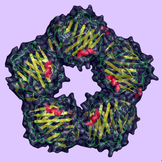
A1AT is a 52 kDa prototypical serine protease inhibitor (serpin) that protects against enzymes released by inflammatory cells, particularly elastase, which is released from neutrophilic granules. A1AT forms covalent bonds with both elastase and trypsin, irreversibly inactivating these proteolytic enzymes.

Alpha 1-antitrypsin deficiency is a hereditary disorder in which inability to inactivate elastase and trypsin allows inflammatory tissue breakdown, causing pulmonary emphysema and hepatic cirrhosis in severe cases.

tags

Labels: , , , , ,

| 0 Guide-Glossary

alpha 2-macroglobulin

Alpha-2 macroglobulin (A2M), α-2 macroglobulin is an an acute phase protein synthesized in response to pro-inflammatory cytokines early in the inflammatory response.

A2M is a large plasma protein produced by the liver, and is a major component of the alpha-2 band in protein electrophoresis. A2M interacts and captures virtually any proteinase whether self or foreign, suggesting a function as a unique "panproteinase inhibitor." A2M also removes the active forms of the gelatinase (MMP-2 and MMP-9) from the circulation, binding to scavenger receptors on the phagocytes. In the mechanism termed "clearance of activated alpha 2-macroglobulin", ACT undergoes Ca(2+)-dependent binding to a member of the low-density lipoprotein receptor supergene family that mediates cellular uptake by endocytosis and delivery to endosomes and lysosomes. Thus, the peptide binding function of A2M allows targeting of biologically active peptides to different cell types expressing the A2M receptor. Complexes internalized through this binding may be dispatched into different pathways of endocytic/lysosomal pathways in a cell type-specific manner.[r]

A2M ratios are increased in nephrotic syndrome when the kidneys lose smaller plasma proteins (proteinuria). A common variant of α2-macroglobulin is associated with increased risk of Alzheimers disease.

 Scavenger Receptors

tags

Labels: , , , , , , , , ,

| 0 Guide-Glossary

CARD domains

Caspase recruitment domains, or CARD domains are found in many proteins, particularly those that evoke inflammatory responses or apoptosis. Proteins with CARD domains include caspases, helicases, kinases, mitochondrial proteins, and other cytoplasmic factors.

CARD domains mediate the formation of large protein complexes through direct interactions between individual caspase recruitment domains. The domains belong to a subclass of protein motif termed the 'death fold', which comprises six to seven antiparallel alpha helices with a hydrophobic core and an outer face composed of charged residues, and which is conserved at least as far as the ced-3 and ced-4 genes in C. elegans. Other 'death fold' motifs in this class are found in the pyrin domain (PYD), death domain (DD), and death effector domain (DED). All function primarily in regulation of apoptosis and inflammatory responses.

The NACHT–leucine-rich repeat (NLR) protein, Ipaf-1 features an N-terminal CARD domain, a nucleotide-binding domain, and C-terminal leucine-rich repeats (LRRs), homologous to those found in Toll-like receptors. Ipaf-1 has a primary role in regulation of proteolytic processing of pro-IL-1β and pro-IL-18 into their mature forms by way of association in a large complex known as the inflammasome.

Ipaf-1 activation by the intracellular bacterium S. typhimurium or other stress signals, causes Ipaf-1 to recruit a CARD-containing adapter termed ASC and caspase-1. This ASC•caspase-1 complex activates caspase-1 and leads to IL-1β and IL-18 maturation.

CARD proteins also participate in recognition of intracellular dsRNA found in a number of viral genomes, including the para-, orthomyxoviridae, and rhabdoviridae. Unlike NLRs, these RIG-I and MDA5 proteins contain twin N-terminal CARD domains plus C-terminal RNA helicase domains that directly interact with and process the double-stranded viral RNA. This processing renders the CARD domains available for interaction with the CARD motif of IPS-1/MAVS/VISA/Cardif, which is a mitochondrion-anchored downstream adaptor.

Because CARD proteins function as regulators of inflammation, the constitutive activation of certain CARD proteins might play a causative role in some inflammatory syndromes.

tags

Labels: , , , ,

| 0 Guide-Glossary

C-reactive protein

C-reactive protein is an acute phase protein produced by the liver within 6 hours of an acute inflammatory stimulus. Levels peak after about 50 hours.

CRP is a pentraxin protein that exhibits calcium dependant ligand binding and a distinctive flattened β-jellyroll structure. (left - image - click to enlarge) CRP is so-named because it reacts with the somatic C polysaccharide of the bacterium Streptococcus pneumoniae. Serum amyloid P component is another pentraxin, acute phase protein.

CRP binds specifically to phosphocholine moieties, conferring a host-defensive role on CRP since phosphocholine is a component of microbial polysaccharides. CRP also binds to ligands exposed on damaged cells.

CRP-phosphocholine-binding:
● activates the classical complement pathway
● acts as an opsonin ligand for phagocytosis
● neutralizes the pro-inflammatory platelet-activating factor (PAF)
● down-regulates polymorphs
CRP
● increases production of tissue-factors by monocytes
● activates smooth muscle K+ ion channels (vasodilator)
● delays apoptosis of neutrophils when the pentameric structure is lost and the molecule exists as a monomer (mCRP)

CRP is mildly elevated in: systemic lupus erythematosus, systemic sclerosis, sermatomyositis, ulcerative colitis, leukaemia, and graft-versus-host disease (GVHD).

The C-reactive protein medical test measures levels of CRP to assess acute inflammation. An association has been demonstrated between sudden cardiac death, peripheral arterial disease and hs-CRP, so serum CRP levels may correlate with cardiovascular disease risk.

tags

Labels: , , , , , , , , ,

| 0 Guide-Glossary

ferritin

Ferritin is an an acute phase protein synthesized in response to pro-inflammatory cytokines early in the inflammatory response.

Ferritin is the chief iron storage protein for both eukaryotes and prokaryotes, and is termed apoferritin when not combined with Fe(3+) ions. The globular ferritin protein complex comprises 24 subunits – hetero-oligomers of light (L) and heavy (H) chains encoded by homologous genes (vertebrates), H chains only in bacteria and plants. Free Fe is toxic, so cells employ ferritin molecules or aggregates (hemosiderin) to form protective Fe-protein complexes. Each ferritin molecule complexes around 4500 (Fe3+)ions.

Ferritin is found in most tissues, particularly in the bone marrow and reticuloendothelial system (or mononuclear phagocytic system). Serum ferritin levels are proportional to iron storage levels in humans and are employed as a part of the work-up for anemia, particurlarly iron-deficiency anemia. However, ferritin levels are increased by acute inflammation, hemochromatosis, malignancy, hyperthyroidism, Still's Disease, or hepatic disease (necrotic hepatocytes), so ferritin levels may be misleading when anemia is concomitant with these conditions.

tags

Labels: , , , , ,

| 0 Guide-Glossary

haptoglobins

Haptoglobins (Hp) are acute phase proteins synthesized in response to pro-inflammatory cytokines early in the inflammatory response.

Haptoglobins are alpha-2-globulins that remove free hemoglobin from plasma by forming a stable complex that aids in recycling of heme iron.

Haptoglobins are elevated in inflammation, malignancies (particularly with metastases to bone), trauma, surgery, steroid or androgen therapy, and diabetes.

tags

Labels: , , , ,

| 0 Guide-Glossary

. . . since 11/21/06